
Page 132 - วารสารกรมการแพทย์แผนไทยฯ ปีที่ 20 ฉบับที่ 1
P. 132
112 วารสารการแพทย์แผนไทยและการแพทย์ ทางเลือก ปีที่ 20 ฉบับที่ 1 มกราคม-เมษายน 2565
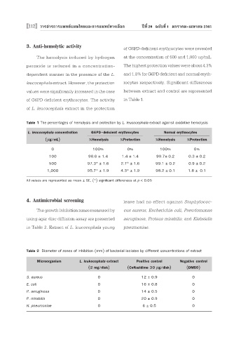
3. Anti-hemolytic activity of G6PD-deficient erythrocytes were revealed
The hemolysis induced by hydrogen at the concentration of 500 and 1,000 ug/mL.
peroxide is reduced in a concentration- The highest protection values were about 4.3%
dependent manner in the presence of the L. and 1.8% for G6PD deficient and normal eryth-
leucocephala extract. However, the protection rocytes respectively. Significant differences
values were significantly increased in the case between extract and control are represented
of G6PD deficient erythrocytes. The activity in Table 1.
of L. leucocephala extract in the protection
Table 1 The percentages of hemolysis and protection by L. leucocephala extract against oxidative hemolysis
L. leucocephala concentration G6PD-deficient erythrocytes Normal erythrocytes
(μg/mL) %Hemolysis %Protection %Hemolysis %Protection
0 100% 0% 100% 0%
100 98.6 ± 1.4 1.4 ± 1.4 99.7± 0.2 0.3 ± 0.2
500 97.3* ± 1.6 2.7* ± 1.6 99.1 ± 0.2 0.9 ± 0.2
1,000 95.7* ± 1.9 4.3* ± 1.9 98.2 ± 0.1 1.8 ± 0.1
All values are represented as mean ± SE, (*) significant differences at p < 0.05
4. Antimicrobial screening leave had no effect against Staphylococ-
The growth inhibition zones measured by cus aureus, Escherichia coli, Pseudomonas
using agar disc diffusion assay are presented aeruginosa, Proteus mirabilis, and Klebsiella
in Table 2. Extract of L. leucocephala young pneumoniae.
Table 2 Diameter of zones of inhibition (mm) of bacterial isolates by different concentrations of extract
Microorganism L. leukocephala extract Positive control Negative control
(2 mg/disk) (Ceftazidime 30 μg/disk) (DMSO)
S. aureus 0 12 ± 0.9 0
E. coli 0 16 ± 0.8 0
P. aeruginosa 0 14 ± 0.5 0
P. mirabilis 0 20 ± 0.9 0
K. pneumoniae 0 6 ± 0.5 0