
Page 86 - วารสารปีที่16ฉบับที่2
P. 86
258
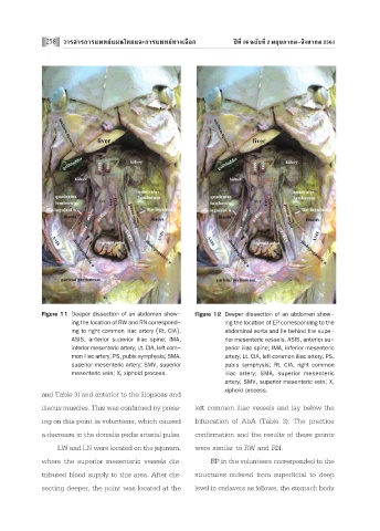
Figure 11 Deeper dissection of an abdomen show- Figure 12 Deeper dissection of an abdomen show-
ing the location of RW and RN correspond- ing the location of EP corresponding to the
ing to right common iliac artery (Rt. CIA). abdominal aorta and lie behind the supe-
ASIS, anterior superior iliac spine; IMA, rior mesenteric vessels. ASIS, anterior su-
inferior mesenteric artery; Lt. CIA, left com- perior iliac spine; IMA, inferior mesenteric
mon iliac artery; PS, pubis symphysis; SMA, artery; Lt. CIA, left common iliac artery; PS,
superior mesenteric artery; SMV, superior pubis symphysis; Rt. CIA, right common
mesenteric vein; X, xiphoid process. iliac artery; SMA, superior mesenteric
artery; SMV, superior mesenteric vein; X,
xiphoid process.
and Table 3) and anterior to the iliopsoas and
iliacus muscles. This was confirmed by press- left common iliac vessels and lay below the
ing on this point in volunteers, which caused bifurcation of AbA (Table 3). The practice
a decrease in the dorsalis pedis arterial pulse. confirmation and the results of these points
LW and LN were located on the jejunum, were similar to RW and RN.
where the superior mesenteric vessels dis- EP in the volunteers corresponded to the
tributed blood supply to this area. After dis- structures ordered from superficial to deep
secting deeper, the point was located at the level in cadavers as follows, the stomach body