
Page 85 - วารสารปีที่16ฉบับที่2
P. 85
257
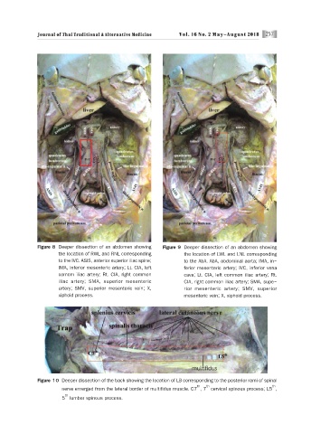
Figure 8 Deeper dissection of an abdomen showing Figure 9 Deeper dissection of an abdomen showing
the location of RWL and RNL corresponding the location of LWL and LNL corresponding
to the IVC. ASIS, anterior superior iliac spine; to the AbA. AbA, abdominal aorta; IMA, in-
IMA, inferior mesenteric artery; Lt. CIA, left ferior mesenteric artery; IVC, inferior vena
comom iliac artery; Rt. CIA, right common cava; Lt. CIA, left common iliac artery; Rt.
iliac artery; SMA, superior mesenteric CIA, right common iliac artery; SMA, supe-
artery; SMV, superior mesenteric vein; X, rior mesenteric artery; SMV, superior
xiphoid process. mesenteric vein; X, xiphoid process.
Figure 10 Deeper dissection of the back showing the location of LB corresponding to the posterior rami of spinal
th th th
nerve emerged from the lateral border of multifidus muscle. C7 , 7 cervical spinous process; L5 ,
th
5 lumbar spinous process.