
Page 52 - journal_13-2_Full
P. 52
130
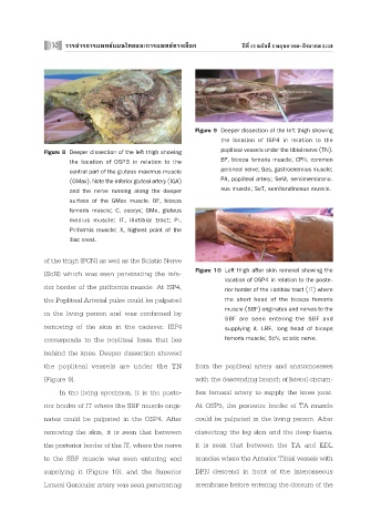
Figure 9 Deeper dissection of the left thigh showing
the location of ISP4 in relation to the
popliteal vessels under the tibial nerve (TN).
Figure 8 Deeper dissection of the left thigh showing
BF, biceps femoris muscle; CPN, common
the location of OSP3 in relation to the
central part of the gluteus maximus muscle peroneal nerve; Gas, gastrocnemius muscle;
(GMax). Note the inferior gluteal artery (IGA) PA, popliteal artery; SeM, semimembrano-
sus muscle; SeT, semitendinosus muscle.
and the nerve running along the deeper
surface of the GMax muscle. BF, biceps
femoris muscle; C, coccyx; GMe, gluteus
medius muscle; IT, iliotibial tract; Pi,
Piriformis muscle; X, highest point of the
iliac crest.
of the thigh (PCN) as well as the Sciatic Nerve
Figure 10 Left thigh after skin removal showing the
(ScN) which was seen penetrating the infe-
location of OSP4 in relation to the poste-
rior border of the piriformis muscle. At ISP4,
rior border of the iliotibial tract (IT) where
the Popliteal Arterial pulse could be palpated the short head of the biceps femoris
muscle (SBF) originates and nerves to the
in the living person and was confirmed by
SBF are seen entering the SBF and
removing of the skin in the cadaver. ISP4 supplying it. LBF, long head of biceps
corresponds to the popliteal fossa that lies femoris muscle; ScN, sciatic nerve.
behind the knee. Deeper dissection showed
the popliteal vessels are under the TN from the popliteal artery and anatomoseses
(Figure 9). with the descending branch of lateral circum-
In the living specimen, it is the poste- flex femoral artery to supply the knee joint.
rior border of IT where the SBF muscle origi- At OSP5, the posterior border of TA muscle
nates could be palpated in the OSP4. After could be palpated in the living person. After
removing the skin, it is seen that between dissecting the leg skin and the deep fascia,
the posterior border of the IT, where the nerve it is seen that between the TA and EDL
to the SBF muscle was seen entering and muscles where the Anterior Tibial vessels with
supplying it (Figure 10), and the Superior DPN descend in front of the interosseous
Lateral Genicular artery was seen penetrating membrane before entering the dorsum of the