
Page 51 - journal_13-2_Full
P. 51
129
Soleus muscles, with the Posterior Tibial ves- men and was confirmed by removing of the
sels and Tibial Nerve (TN) then seen under thigh skin in the cadaver. OSP2 is in the vi-
the Flexor Digitorum Longus (FDL) muscle. cinity of the posteroinferior border of the ASIS
The lateral border of tibia could be pal- and was at the fleshy part of the TFL muscle.
pated adjacent NP1 and NP2 in the living The Superior Gluteal vessels and SGN were
person. After removing the skin, it was found seen entering the TFL and supplying it. When
that both NP1 and NP2 are located between dissecting deeper, the Iliofemoral ligament
the origins of the TA and EDL muscles, with was found to overly the head of the femur in
the Anterior Tibial vessels seen penetrating the acetabulum (Figure 7). At OSP3, the cen-
through the openings in the interosseous tral part of GMax muscle could be palpated
membrane at NP1, and DPN ramifying to in the living person and was confirmed by
supply the muscles at NP2 (Figure 1). At removing of the thigh skin in the cadaver.
TAOP, the Femoral Arterial pulse could be OSP3 associates with the Inferior Gluteal ves-
palpated in the living person and was con- sels and Inferior Gluteal Nerve (IGN), which
firmed by removing the thigh skin in the ca- were seen entering the central part of the
daver. TAOP is in the superior part of the GMax and supplying it (Figure 8). OSP3 also
femoral triangle where the Femoral vessels corresponds to the Posterior Cutaneous Nerve
and FN disappear into the adductor canal
through the apex of femoral triangle.
The posterior border of IT could be pal-
pated adjacent OSP1 in the living person.
Removal of the skin showed that OSP1 corre-
sponds to the anterosuperior border of the
GMax muscle at a location adjacent to the
posterior border of the IT. It was also superfi-
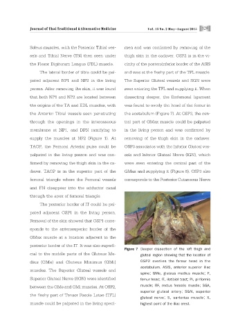
Figure 7 Deeper dissection of the left thigh and
cial to the middle parts of the Gluteus Me- gluteal region showing that the location of
dius (GMe) and Gluteus Minimus (GMi) OSP2 overlies the femur head in the
acetabulum. ASIS, anterior superior iliac
muscles. The Superior Gluteal vessels and
spine; GMe, gluteus medius muscle; F,
Superior Gluteal Nerve (SGN) were identified femur head; IT, ilotibial tract; Pi, piriformis
between the GMe and GMi muscles. At OSP2, muscle; RF, rectus femoris muscle; SGA,
superior gluteal artery; SGN, superior
the fleshy part of Tensor Fascia Latae (TFL)
gluteal nerve; S, sartorius muscle; X,
muscle could be palpated in the living speci- highest point of the iliac crest.