
Page 91 - วารสารปีที่15ฉบับที่2
P. 91
216
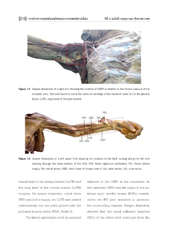
Figure 15 Deeper dissection of a right arm showing the location of SSP5 in relation to the fibrous capsule of the
shoulder joint. This was found to overly the articular cartilage of the humeral head (H) in the glenoid
fossa. LoTB, long head of tricepbs brachii.
Figure 16 Deeper dissection of a left upper limb showing the location of the BLIF running along the MN and
passing through the deep surface of the FDS. FDP, flexor digitorum profundus; FPL, flexor policis
longus; RA, radial artery; SBB, short head of biceps brachii; UA, ulnar artery; UN, ulnar nerve.
lateral head of the triceps brachii (LaTB) and adjacent to the OSP3 in the volunteers. In
the long head of the triceps brachii (LoTB) the cadavers, OSP3 was the origin of the ex-
muscles. On deeper dissection, radial nerve tensor carpi radialis longus (ECRL) muscle,
(RN) ramified to supply the LoTB and passed where the RN gave branches to innervate
inferolaterally into the radial groove with the the surrounding muscles. Deeper dissection
profunda brachii artery (PBA; Table 2). showed that the radial collateral ligament
The lateral epicondyle could be palpated (RCL) of the elbow joint continued from the