
Page 85 - วารสารปีที่15ฉบับที่2
P. 85
210
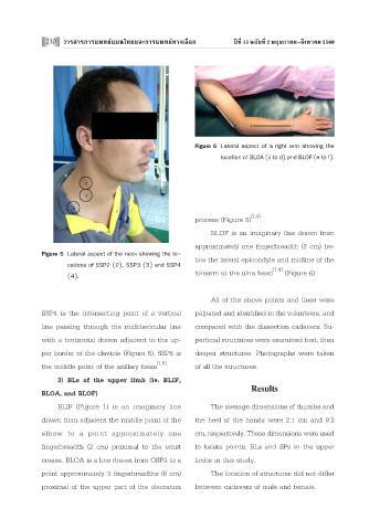
Figure 6 Lateral aspect of a right arm showing the
location of BLOA (c to d) and BLOF (e to f).
[1,4]
process (Figure 6) .
BLOF is an imaginary line drawn from
approximately one fingerbreadth (2 cm) be-
Figure 5 Lateral aspect of the neck showing the lo-
low the lateral epicondyle and midline of the
cations of SSP2 (2), SSP3 (3) and SSP4
[1,4]
forearm to the ulna head (Figure 6).
(4).
All of the above points and lines were
SSP4 is the intersecting point of a vertical palpated and identified in the volunteers, and
line passing through the midclavicular line compared with the dissection cadavers. Su-
with a horizontal drawn adjacent to the up- perficial structures were examined first, then
per border of the clavicle (Figure 5). SSP5 is deeper structures. Photographs were taken
[1,5]
the middle point of the axillary fossa . of all the structures.
3) BLs of the upper limb (ie. BLIF,
Results
BLOA, and BLOF)
BLIF (Figure 1) is an imaginary line The average dimensions of thumbs and
drawn from adjacent the middle point of the the heel of the hands were 2.1 cm and 9.2
elbow to a point approximately one cm, respectively. These dimensions were used
fingerbreadth (2 cm) proximal to the wrist to locate points, BLs and SPs in the upper
crease. BLOA is a line drawn from OSP2 to a limbs in this study.
point approximately 3 fingerbreadths (6 cm) The location of structures did not differ
proximal of the upper part of the olecranon between cadavers of male and female.